Chemistry that draws. Measured, recorded, reproducible.
Two substances react and spread across a grid. Nobody draws the shapes; they emerge from the chemistry. Every sample keeps its recipe, seed and generation on file, so it can be grown again exactly.
- F
- 0.0545
- k
- 0.0620
- Seed
- 2026
- Grid
- 320 × 200
- Δt
- 1.0
- Generation
- 0
Six recipes in culture
Same chemistry, different parameters. Feed rate F and kill rate k decide whether the grid becomes a labyrinth, spots or foam. These slides are growing right now; open any of them in the Studio to keep cultivating it.
The Gray–Scott model
The mechanism Alan Turing proposed in 1952 to explain stripes and spots in nature, in the simplified form John Pearson studied in 1993.
∂v/∂t = Dv∇²v + uv² − (F + k)v
u = substrate · v = activator · Du = 1 · Dv = 0.5 · 9-point Laplacian · periodic boundaries
| 01 | Seed Drops of activator enter the grid through the brush, a word, a photo or a video. |
| 02 | React The activator multiplies by consuming substrate. F refills the substrate; k removes the activator. |
| 03 | Diffuse The substrate spreads twice as fast as the activator. Shapes are born from that imbalance. |
From the cell to the grid
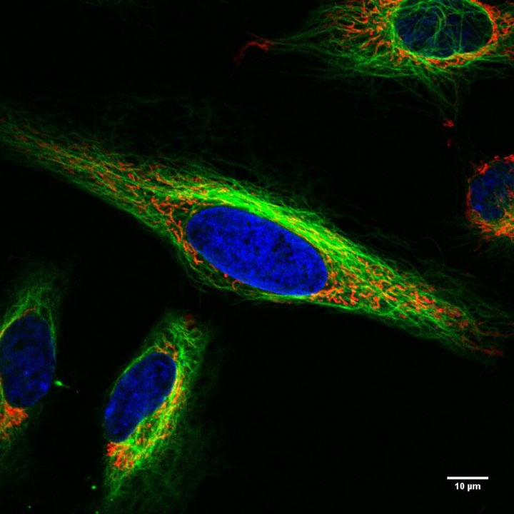
Cultivate patterns
Brush, walls, eight recipes, palettes, high-resolution PNG and video of the growth.
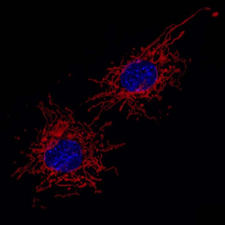
Images become texture
Dark and light areas grow with different recipes, motion leaves trails, and the source reappears made of pattern.
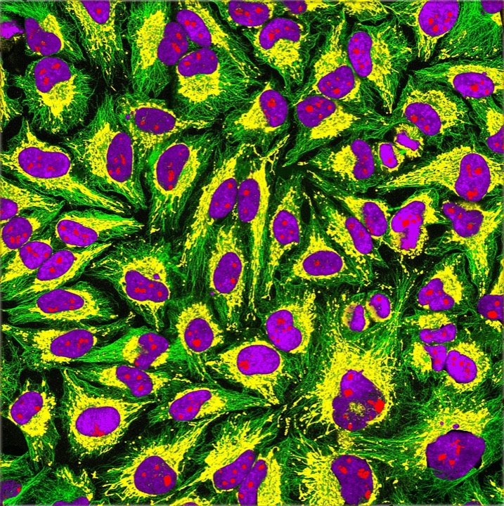
Virtual cell
Reactions, compartments, and deterministic, stochastic and spatial simulations of cell biology.
A laboratory and a studio in one instrument
ORGANELA started as a virtual cell, inspired by Virtual Cell (VCell) from the Center for Cell Analysis and Modeling at UConn Health, where you describe reactions and the software takes care of the math.
The same math that simulates proteins inside a cell also makes images. Every sample on this page is a real simulation, computed on your machine and reproducible from its seed.
Turing, A. M. (1952). The Chemical Basis of Morphogenesis. Pearson, J. E. (1993). Complex Patterns in a Simple System. Nine-point Laplacian after Karl Sims.
Microscopy photographs: Wikimedia Commons, under the Creative Commons licenses listed on each file: HeLa, MEF, HeLa-Tubulin.


